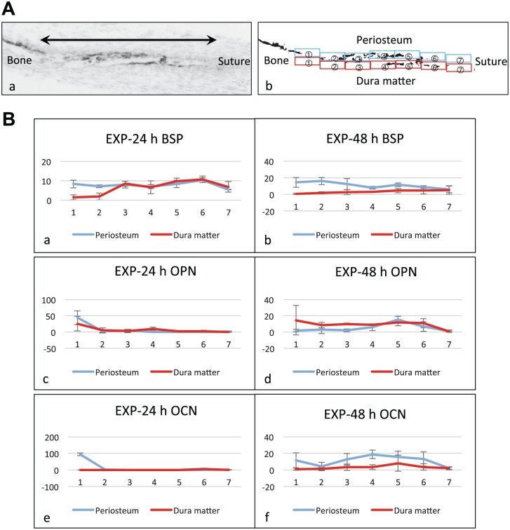
Figure 4.
Quantification of expression pattern of bone matrix proteins. (A) Indication of the measurement areas. (a) A gray scale image of a cryosection hybridized with the antisense probe for bone matrix protein. The bone surface areas from the calcified bone margin (left side: Bone) to the tip of the osteoid (right side: Suture), indicated by the double headed arrow in the gray scale image, were equally divided into seven measurement areas. (b) A binarized image of (a). The measurement areas were numbered from 1 to 7 in the direction from bone to suture side on each of the periosteum side (blue squares) and the dura mater side (red squares). The percentage of the reaction-positive areas for in situ hybridization (ISH) in each measurement area was calculated on binarized images. (B) Graphs showing the quantification results of ISH for the stretched experimental group (EXP) at 24 hr and 48 hr (n=3). The vertical axis and horizontal axis correspond to the percentage of the reaction-positive areas and to the numbers of the measurement areas, respectively. Error bars: Standard deviation. (a and b) Reaction-positive areas for bone sialoprotein (BSP) expression. In EXP-24 h group, they were observed overall both on the periosteum side and the dura matter side (a). In EXP-48 h group, the reaction-positive areas decreased on the dura mater side (b). (c and d) Reaction-positive areas for osteopontin (OPN) expression. In EXP-24 h group, they were observed partially on the dura matter side but were hardly detected on the periosteal side (c). In EXP-48 h group, the reaction-positive areas were observed overall on the dura mater side and partially on the periosteum side (d). (e and f) Reaction-positive areas for osteocalcin (OCN) expression. In EXP-24 h group, OCN was rarely expressed in the newly formed area (e). However, in EXP-48 h group, the reaction-positive areas increased on the periosteum side (f). Particularly, osteoblasts located in the areas 3 and 4 of the periosteum side in EXP-48 h group mainly expressed OCN rather than OPN (d and f).

