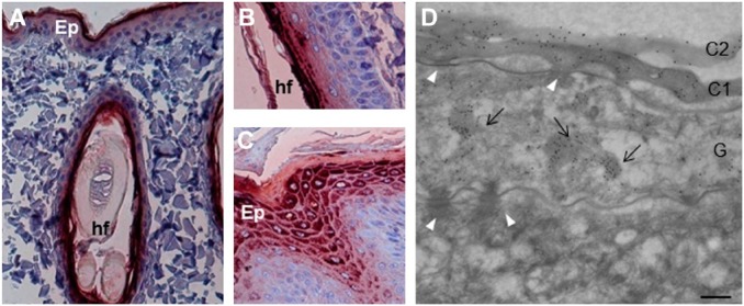
Figure 2.
Localization of filaggrin in the skin of healthy dogs. (A) The mAbs of the AHF family were analyzed by immunohistochemistry on sections of dog skin, from back and paw pad. AHF10, 13, 23, and 27 displayed the same pattern of reactivity, but only the data for AHF10 are shown. (A–C) AHF10 stains the upper interfollicular epidermis (A) and the intrafollicular epidermis (B) of the back skin, and the upper epidermis of paw pad (C). (D) The reactivity of AHF10 was further analyzed using immunoelectron microscopy. The mAb labels the first two layers of corneocytes (C1 and C2) and the keratohyalin granules (arrows) in a granular (G) keratinocyte. The spinous (S) keratinocytes as well as desmosomes (white arrowheads) are not labeled. Scale bar = 30 µm (A and C), 25 µm (B) and 0.25 µm (D). Abbreviations: AHF, anti-human filaggrin; Ep, epidermis; hf, hair follicle.

