Figure 4.
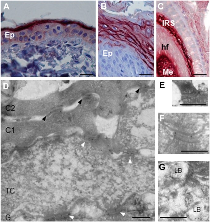
Localization of corneodesmosin in the skin of healthy dogs. (A–C) Using immunohistochemistry, G36-19 mAb stains the upper interfollicular epidermis (Ep) of back skin (A), the upper epidermis of paw pad (B) and both the inner root sheath (IRS) and medulla (Me) of hair follicles (hf, C). (D–G) G36-19 was further used in immunoelectron microscopy. (D) The reactivity is localized in the extracellular part of corneodesmosomes (black arrowheads) in the lower cornified layer, and of desmosomes (white arrowheads) in transitional cells (TC) and granular keratinocytes (G). The first two layers of corneocytes (C1 and C2) are shown. (E, F) Enlargements of desmosomes in transitional and granular cells. (G) lamellar bodies (LB) of granular cells are also labeled. Scale bar = 30 µm (A–C), 0.3 µm (D–G).

