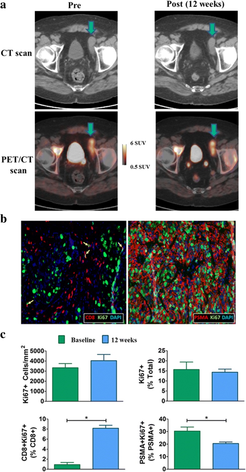
Fig. 4.

a Axial CT and PET/CT slices with a metastatic tumor indicated. At week 12 this patient had experienced diminished PSA and RECIST measurements but increased tumor FLT uptake. By week 16, this patient was found to have progressive disease with marked increases in tumor size and PSA. b Immunofluorescence images show representative FFPE sections taken from the week 12 biopsy of the tumor indicated in part (a). The left immunofluorescence image shows proliferating T cells (Ki67 + CD8+; yellow arrows) and the right image shows proliferating tumor cells (Ki67 + PSMA+). c Quantification of the immunofluorescence images from the tumor indicated in part (a). The top row shows changes in the number of proliferating cells per unit area (left) and changes in the percentage of proliferating cells (right). The bottom row shows percent changes in proliferating CD8+ T cells (left) and proliferating PSMA+ tumor cells (right). *P-value less than 0.05
