Fig. 1.
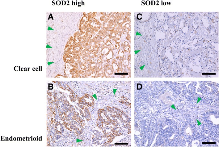
Examples of mitochondrial superoxide dismutase (SOD2) expression in endometriosis-associated ovarian cancers on immunohistochemical analysis. Cases subjected to immunohistochemical analysis for detection of mitochondrial superoxide dismutase (SOD2) expression in clear cell carcinoma and endometrioid carcinoma are shown in (a, c) and (b, d), respectively. High and low mitochondrial superoxide dismutase (SOD2) expression levels are demonstrated in (a, b) and (c, d), respectively. Green arrowheads indicate stromal cell areas, where SOD2 was subtly expressed and used as internal controls. The scale bars correspond to 100 μm

