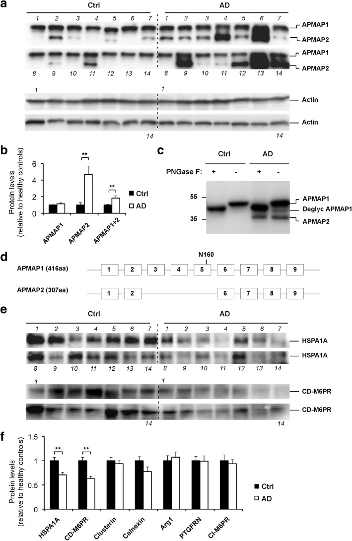
Fig. 5.
Increased alternative splicing of APMAP and reduced HSPA1A and CD-M6PR in AD brains. a Increased alternative splicing variant APMAP2 in AD brains, as estimated by Western blot analysis of APMAP1 and APMAP2 in cortical lysates of 14 control brains and 14 neuropathologically verified AD brains. Detailed demographic and diagnostic features of the human brain samples are provided in Table 1. Actin served as a loading control. b Densitometric analysis of the APMAP1 and APMAP2 Western blot bands in (a). Student’s t-test with mean ± SEM, **P < 0.01. c Denatured cortical lysates of control and AD brains treated in the presence (+) or absence (-) of PNGase. d Schematic representation of the exons and introns of APMAP1 and APMAP2. The predicted glycosylation site in exon 5 at position N160 is shown. e Reduced HSPA1A and CD-M6PR levels in AD brains, as estimated by Western blot analysis in the same samples as in (a). f Densitometric analysis of HSPA1A and CD-M6PR (e) and other APMAP-interactomers Additional file 1 Figure S9 Western blot bands. Student’s t-test with mean ± SEM, **P < 0.01

