
Figure 2: CD8 T cells that express NKG2A belong to CD103+ early effector tissue-resident cells.
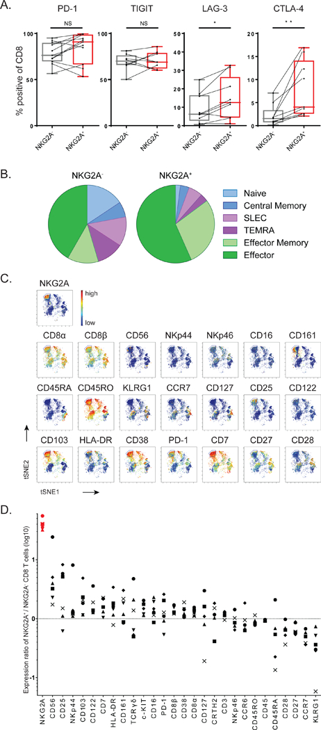
(A) Co-expression flow cytometry analysis of inhibitory receptors on CD8 TIL in HNSCC biopsies. Paired Student’s t-test. (B) T cell differentiation stage of NKG2A-positive CD8 TIL was determined by combined expression of these markers: naive (CCR7+ CD127-), central memory (CCR7+ CD127+), SLEC (‘short lived effector cells’: CCR7- CD45RO+ KLRG1+), TEMRA (‘CD45RA+ effectors’: CCR7’CD45RO’), effector memory (CCR7- CD45RO+ KLRG1- CD127+) and effector (CCR7- CD45RO+ KLRG1- CD127-). (C) Density viSNE plot from CyTOF data on pre-gated CD3+CD8+ TIL. (D) Ratios of marker expression (based on the Mean Signal Intensity) on NKG2A+ versus NKG2A- CD8 T cells in cervical carcinomas (n=6). Each symbol represents an individual tumor sample. See also Figures S1 and S2.