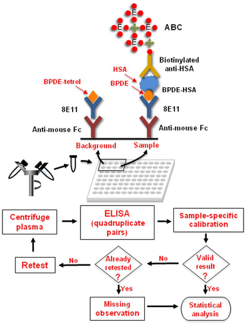
Figure 1.
Sandwich ELISA for direct measurement of BPDE-HSA in plasma. After centrifugation, plasma is mixed with reagents and loaded into pairs of wells in a pre-coated plate. Because the anti-HSA detection antibody responds to both BPDE-HSA (about one nM in plasma) and unadducted HSA (about one mM in plasma), a high background signal is created from amplification of non-specifically bound HSA on well surfaces. To overcome this problem, paired measurements (‘background’ and ‘sample’) are obtained for each plasma sample in quadruplicate and the signals are deconvoluted and calibrated by a sample-specific calibration method (Figure 2). Background wells are treated with BPDE tetrols to deactivate the capture antibody (8E11) and thereby to produce matched controls. To minimize measurement errors, each result is screened for validity using predetermined rules. Valid samples are used for statistical analyses and invalid samples are treated as missing observations.

