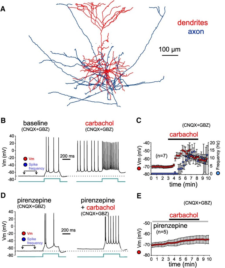
Figure 10.
Effects of carbachol on the membrane potential and excitability of L3-6 PNs, tested in the presence of synaptic receptor blockers (CNQX and gabazine). A, Reconstruction of the dendritic tree of a layer 5 PN for which the effects of carbachol were assessed during current clamp recordings. B, Examples of membrane potential (Vm) recorded from a layer 5 PN before (baseline) and during application of 20 µM carbachol (carbachol), in the presence of synaptic receptor blockers CNQX and gabazine (GBZ). The time window for measurements of Vm (minimum value within the window) and of spike frequency (independent of injection of current steps) is illustrated in the baseline recording example. The injected current step had identical amplitude (70 pA) in baseline and carbachol conditions. Note that during the depolarization induced by carbachol, action potential amplitude was typically reduced. C, Plot illustrating the time course of changes in Vm (red) and in spike frequency (blue) measured during the time window indicated in B. Carbachol depolarized the membrane potential significantly by 2 min of carbachol application (baseline: –71 ± 2 mV, carbachol: –52 ± 5 mV, n = 7, t(6) = 5.04, p = 0.0023, paired t test). Note that for several PNs the strong depolarization by carbachol produced depolarization block of action potential firing, which was prevented by hyperpolarizing current injection (see text in Results). Measurements obtained during hyperpolarizing current injection were not included in this time-course plot. D, Examples of Vm recordings from a layer 5 PN in the continuous presence of the M1 mAChR antagonist pirenzepine (1 µM) before (pirenzepine) and during application of 20 µM carbachol (pirenzepine+carbachol). Vm and spike frequency measurements were performed as indicated in C. The injected current step had identical amplitude (80 pA) in baseline and carbachol conditions. Note that in the presence of pirenzepine, the carbachol-induced depolarization was subthreshold and did not evoke spikes independent of the injected current steps. E, Plot illustrating the time course of changes in Vm. In the presence of pirenzepine, carbachol depolarized the membrane potential, but the depolarization was small and remained below action potential threshold. The depolarization was not significant by 2 min of application but peaked at significant levels by 5 min (baseline: –69 ± 2 mV, 2 min pirenzepine+carbachol: –67 ± 3 mV. 5 min pirenzepine+carbachol: –65 ± 3 mV, n = 5, F(2,8) = 8.555, p = 0.0103, one-way RM ANOVA; baseline vs 2 min: p = 0.239, baseline vs 5 min: p = 0.0034, Dunnett's post hoc tests).

