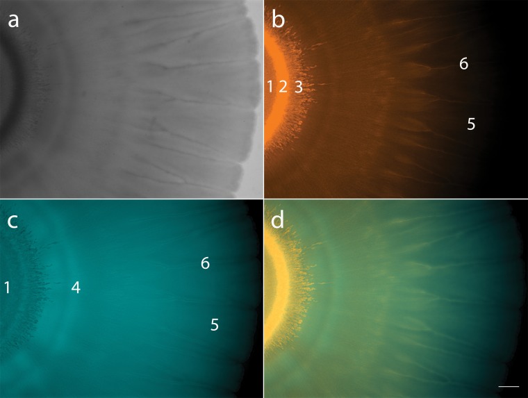
FIG 3.
Mixed colony of fluorescently tagged P. fluorescens Pf0-1 (Pf0-ecfp) and Pedobacter sp. V48 (V48-dsRed). (a) Coculture colony viewed with white light. (b) Coculture imaged using DsRed filter (filter set 43 HE), pseudocolored in orange, showing V48-dsRed distribution throughout the colony. (c) Coculture imaged using CFP filter (filter set 47 HE), pseudocolored in turquoise, showing Pf0-ecfp distribution throughout the colony. (d) Merged images of DsRed and CFP filters. Numbers in panels b and c indicate six zones of distinct patterns: 1, point of inoculation; 2, coffee ring; 3, starburst; 4, P. fluorescens ring; 5, petals; 6, veins. Colonies imaged at ×7 magnification; scale bar represents 1 mm. Colony imaged 144 h after inoculation.

