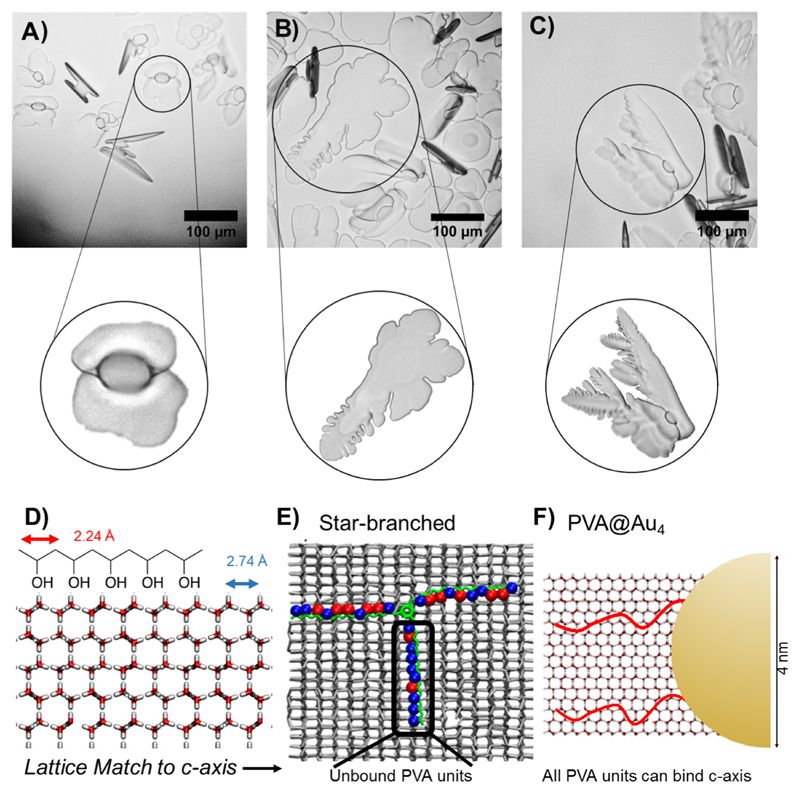
Figure 4.
Ice shaping and binding. Cryomicroscopy images in 45% sucrose with zoomed image of crystals of interest. (A) No additive, −6 °C. (B) PVA140 0.32 mg mL−1, −5.5 °C. (C) PVA140-Au4 0.32 mg mL−1, −4 °C. (D) Pattern matching of PVA to the prism plane of ice. (E) (adapted with permission from J. Phys. Chem. C 2017, 121, 26949–26957) shows 3-arm PVA binding to ice, with red circles indicating bound hydroxyls and blue unbound. Highlighted arm has little binding due to being misaligned with lattice. (F) Schematic of gold particle highlighting low density of grafted polymer, does not constrain and hence can find prismatic faces to bind. Scale bars = 100 μm.

