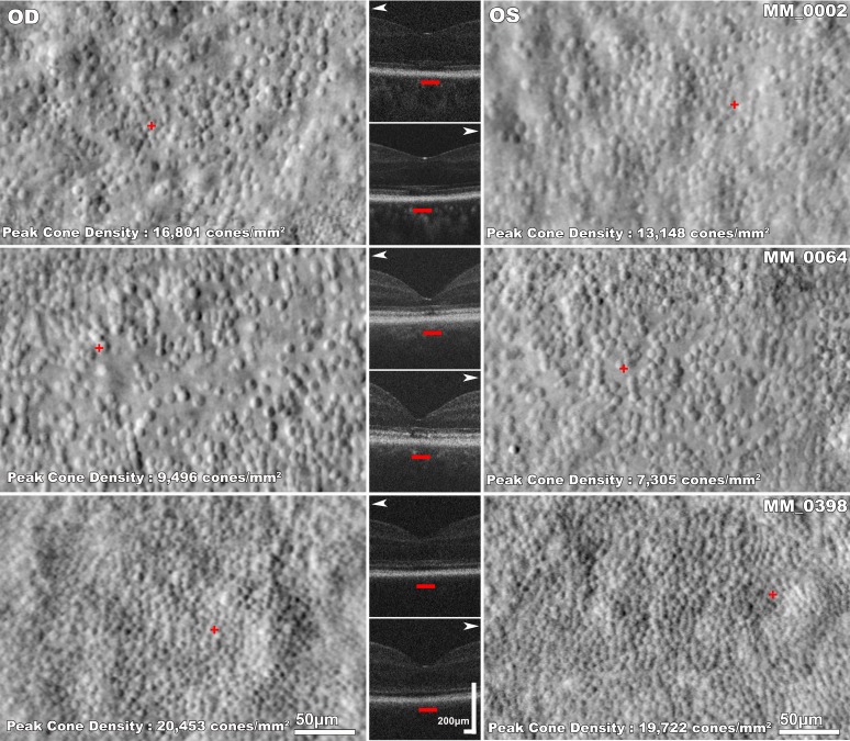
Figure 4.
Disease symmetry in CNGA3-associated ACHM. Left and right column: split-detection AOSLO images of the right and left eye respectively of three subjects with CNGA3-ACHM. Middle column: horizontal transfoveal OCT scans of the same subjects. White arrowheads point to the respective AOSLO image. The red horizontal bars mark the 300-μm area represented in the AOSLO images. There is substantial variability in foveal photoreceptor mosaic between the three subjects. There are structural similarities in the mosaic pattern of the right and left eyes of each subject; MM_0002 and MM_0064 have noncontiguous sparse foveal mosaics and MM_0398 has a relatively contiguous mosaic. MM_0002 has OCT grade 4 bilaterally with foveal hypoplasia. MM_0064 and MM_0398 have EZ grade 2 bilaterally.

