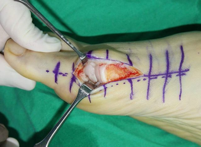
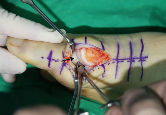
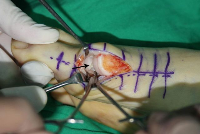
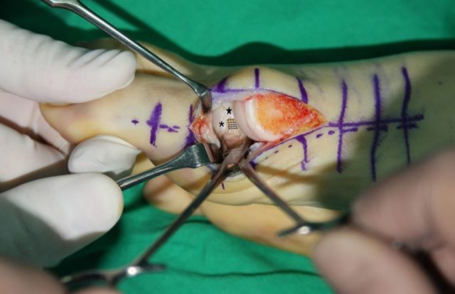
Figs. 3-A through 3-E Anatomical structures to be released through the medial transarticular approach.
Fig. 3-A.
The first metatarsophalangeal joint is distracted distally with use of two vein retractors and application of manual traction to the great toe to allow clear visualization of the lateral soft-tissue structures.
Fig. 3-B.
The lateral joint capsule (arrow) is visualized through the metatarsophalangeal joint, which is distracted with use of a mosquito clamp.
Fig. 3-C.
After the lateral joint capsule is released with use of a number-15 blade, the adductor hallucis tendon (arrow) is observed.
Fig. 3-D.
The adductor hallucis tendon is released completely. The dotted lines show the released adductor hallucis, the star indicates the remnant of the dorsolateral joint capsule, and the asterisk marks the proximal phalangeal portion of the released lateral joint capsule.
Fig. 3-E.
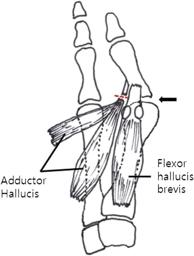
Illustration showing the conjoined tendon of the adductor hallucis tendon transected from its insertion into the base of the proximal phalanx.

