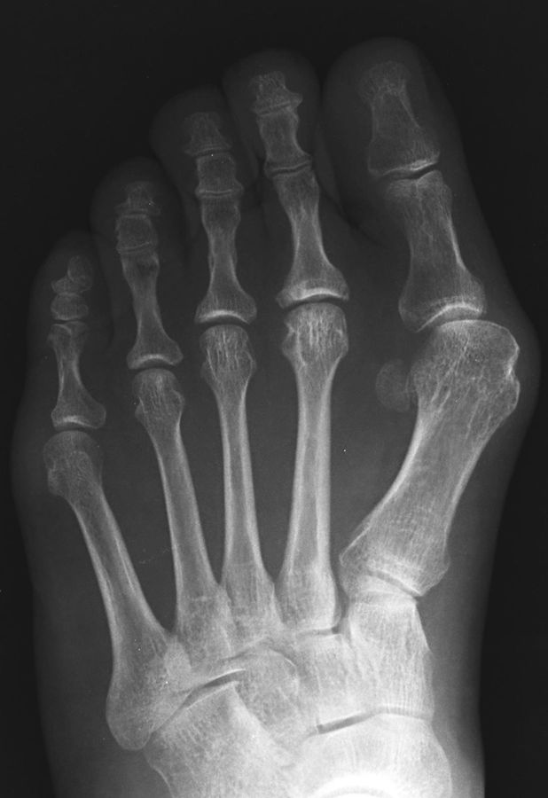
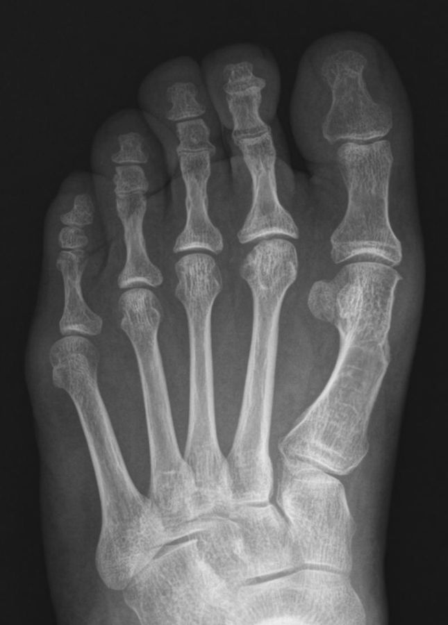
Figs. 9-A through 9-E Clinical photographs and radiographs of the left foot of a sixty-year-old woman who underwent distal chevron osteotomy with a distal soft-tissue procedure through the medial transarticular approach for severe hallux valgus deformity.
Fig. 9-A.
The preoperative hallux valgus angle was 38°, the first-second intermetatarsal angle was 19°, and the tibial sesamoid position was grade 3.
Fig. 9-B.
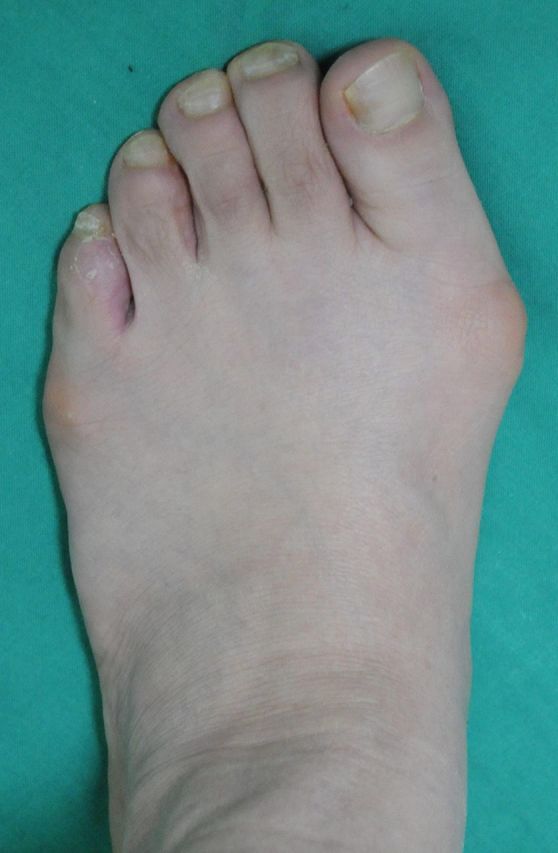
Preoperative clinical photograph.
Fig. 9-C.
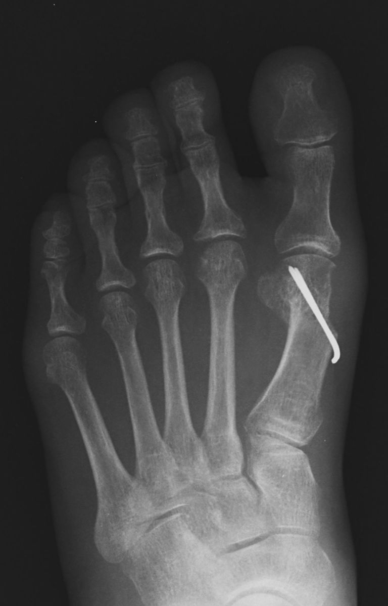
Postoperative anteroposterior radiograph.
Fig. 9-D.
The hallux valgus angle was 10°, the first-second intermetatarsal angle was 9°, and the tibial sesamoid position was grade 2 at the time of final follow-up.
Fig. 9-E.
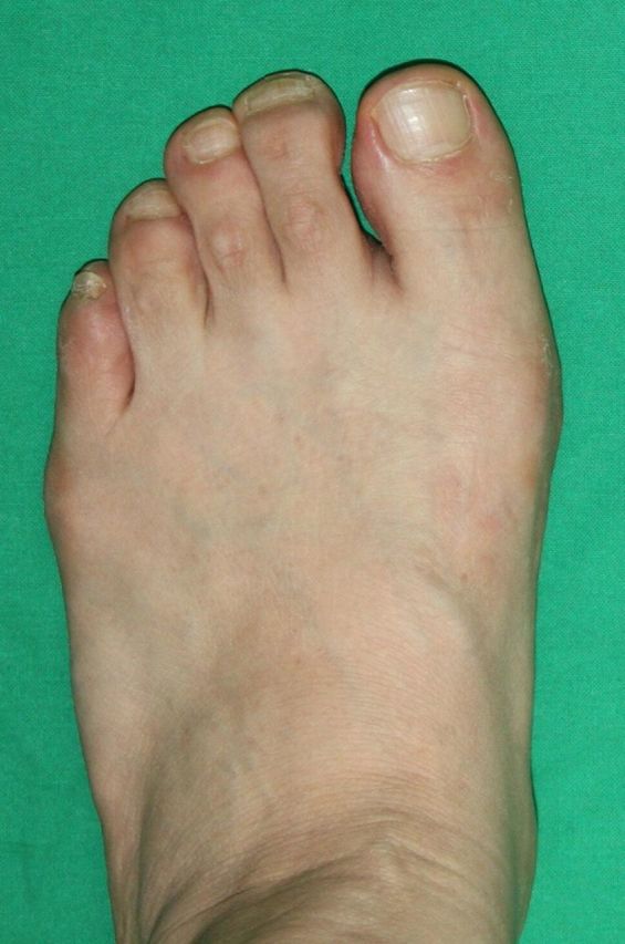
Clinical photograph at the time of final follow-up.

