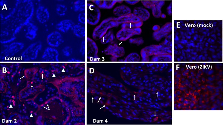
Fig 10.
Pan flavivirus immunofluorescence (IF; Red: flavivirus; Blue: DAPI) staining in placenta from control (A), and three placenta from ZIKV infected dams that ZIKV RNA+ in placenta (B, Dam 3, 14 dpi; C, Dam 3, 14 dpi; D, Dam 4, 21 dpi). The most intense IF was observed in the placenta of Dam 2 (B) in which there was fetal death. ZIKV IF was observed in syncytiotrophoblast (arrows) in Dam 2 and 3. Arrowheads in B indicate IF in the villous cores which could be either stromal or endothelial cells. A less intense IF pattern was observed in Dam 4 (D) in the overlying syncytiotrophoblast which is covering the underlying cytotrophoblasts, despite widespread vertical transfer of ZIKV to the fetus.

