Figure 1.
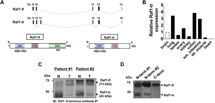
Alternative splicing results in a Raf1-tr protein isoform: A) Exon/intron map of Raf1-fl and the identified splice isoform (Raf1-tr) (top). New exon in Raf1-tr is highlighted in red. Protein product of Raf1-fl (bottom left) showing conserved regions containing a Ras-binding domain and cysteine-rich domain (blue), serine-threonine–rich domain (red), and kinase domain (green). Raf1-tr protein product (bottom right) is truncated and has all conserved regions except for the kinase domain. B) Measurement of Raf1-tr gene expression in a panel of human tissues. The gene of interest was normalized to GAPDH expression and represented as fold change relative to brain tissue. C) Western blot of Raf1 in matched normal (N) and adenocarcinoma colon (T) lysate showing that Raf1-tr protein is differentially expressed in normal and colon tumor tissue. D) Probing for Raf1 with 2 unique N-terminal targeted antibodies demonstrates a 73 kDa Raf1-fl species and a 43 kDa Raf1-tr species in HEK cell lysate. Probing for Raf1 with a C-terminal targeted antibody detects only the 73 kDa Raf1-fl species.

