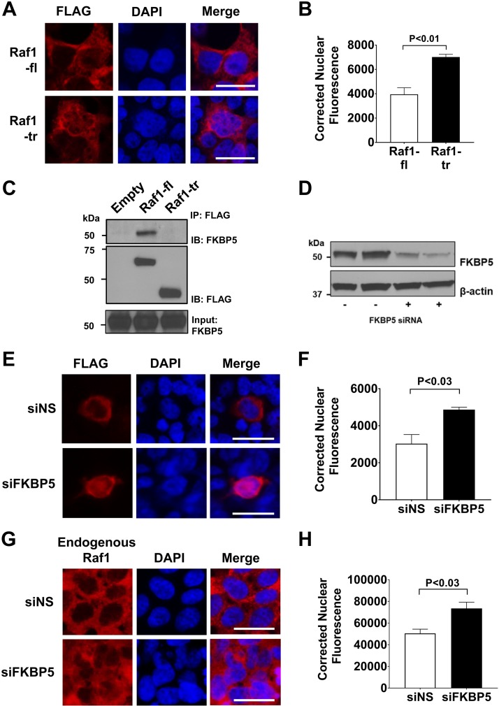
Figure 4.
Truncated Raf1 has increased nuclear localization: A) Measurement of cellular localization of FLAG tagged Raf1-fl (top) or Raf1-tr (bottom) that was expressed in HEK cells. FLAG immunofluorescence is shown in red with nuclei (blue) counterstained with DAPI. Scale bars, 25 µm. Images are representative of experiments run in triplicate. B) Quantitation of nuclear Raf1 localization by corrected nuclear fluorescence calculation. A minimum of 280 cells per group were analyzed. C) FLAG-tagged Raf1 (Raf1-fl) or Raf1-tr was expressed in HEK cells with subsequent FLAG immunoprecipitation and Western blot for FKBP5. D) Western blot of FKBP5 after siRNA knockdown of FKBP5 in HEK cells. β-actin was used as a loading control. E) Measurement of FLAG-tagged Raf1-fl localization (red) after control nonspecific siRNA (top, siNS) or FKBP5 siRNA (bottom, siFKBP5) by immunofluorescence. Nuclei (blue) were counterstained with DAPI. Scale bar, 25 µm. Images are representative of experiments run in triplicate. F) Quantitation of nuclear Raf1-fl localization by corrected nuclear fluorescence calculation. A minimum of 200 cells per group were analyzed. G) Measurement of endogenous Raf1 localization (red) after control nonspecific siRNA (top, siNS) or FKBP5 siRNA (bottom, siFKBP5) by immunofluorescence. Nuclei (blue) were counterstained with DAPI. Scale bar, 25 µm. Images are representative of experiments run in triplicate. H) Quantitation of nuclear Raf1 localization by corrected nuclear fluorescence calculation. A minimum of 300 cells per group were analyzed. The results are shown as means ± sem.

