Figure 6.
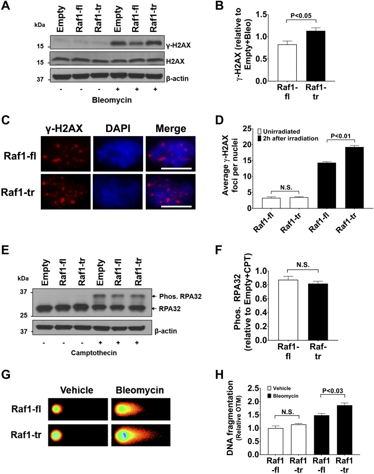
Raf1-tr increases DNA damage: A) Western blot of γ-H2AX and H2AX in HEK cells expressing empty vectorRaf1-fl, or Raf1-tr exposed to vehicle (−) or 0.05 mg/ml bleomycin (+) for 2 h. β-actin was used as a loading control. Blot is representative of experiments run in triplicate. B) Quantitation of γ-H2AX normalized to empty vector exposed to bleomycin (Bleo). C) HEK cells expressing Raf-fl or Raf1-tr were irradiated with 1 Gy of radiation and allowed to recover in complete medium for 2 h. γ-H2AX immunofluorescence is shown in red with nuclei (blue) counterstained with DAPI. Scale bars, 10 µm. Images are representative of experiments run in triplicate. D) Quantitation of γ-H2AX foci. A minimum of 180 nuclei per group were analyzed. E) Western blot of RPA32 phosphorylation in HEK cells expressing empty vector, Raf1-fl, or Raf1-tr exposed to vehicle (−) or 1 µM camptothecin (+) for 1 h. Exposure to camptothecin results in a non-phosphorylated RPA32 band (lower) and a slower migrating phosphorylated band (upper). β-actin was used as a loading control. Blot is representative of experiments run in triplicate. F) Quantitation of RPA32 phosphorylation normalized to empty vector exposed to camptothecin (CPT). G) Neutral comet assay of nuclei from HEK cells expressing Raf1-fl or Raf1-tr exposed to vehicle or 0.05 mg/ml bleomycin for 30 min. Images are representative of experiments run in triplicate. H) Quantitation of DNA fragmentation by Olive tail moment (OTM). A minimum of 200 nuclei per group were analyzed. The results are shown as means ± sem. N.S., not significant.

