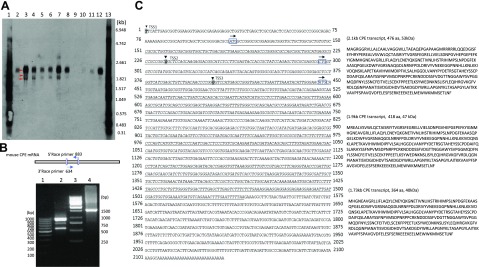
Figure 1.
Identification of CPE transcript variants in embryonic mouse brain. A) Northern blot analysis of RNA isolated from the head of embryos at various developmental stages (E8.5, E10.5, E12.5, E14.5, and E16.5) and from P1 cortex, P1 hippocampus, and adult mouse hippocampus, heart, lung, and liver. The membrane was hybridized to a digoxigenin (DIG)-labeled CPE probe. Lanes 1 and 13, DIG-labeled RNA marker; lane 2, E8.5 hippocampus; lane 3, E10.5; lane 4, E12.5; lane 5, E14.5; lane 6, E16.5; lane 7, P1 cortex; lane 8, P1 hippocampus; lane 9, adult liver; lane 10, adult lung; lane 11, adult heart; and lane 12, adult hippocampus. Three CPE transcripts (2.1, 1.9, and 1.73 kb) are detected as indicated by the arrows. B, Upper) Schematic illustration of the relationships of primers used in the RACE PCRs. Numbers refer to the position of the primers. B, Lower) 5′/3′-RACE PCRs were performed with mRNA isolated from E14.5 mouse brain. Lane 1, 100-bp DNA marker; lane 2, 5′-883 RACE PCR; lane 3, 1-kb DNA marker; and lane 4, 3′-634 RACE PCR. C) The nucleotide sequences of the full-length mRNAs and the deduced proteins of mouse CPE-WT and the 2 variants. Left) Nucleotide sequences of the full-length mRNAs, boldface italic, shaded letters indicated with arrowheads are transcription starting sites of CPE-WT, 1.9 and 1.73 kb CPE transcripts. Arrows above boxed letters indicate potential translation start sites at 110, 296, and 446 nt used by CPE-WT, 1.9 and 1.73 kb CPE transcript variant, respectively. Right) Deduced amino acid sequences of the 3 CPE transcripts.

