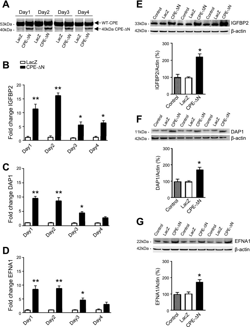
Figure 4.
The 40-kDa CPE-∆N up-regulates expression of IGFBP2, DAP1, and EFNA1 genes in HT22 neurons. A) Western blot showing 40-kDa CPE-∆N expression and LacZ control in HT22 neurons transduced with 10 MOI virus. B–D) Bar graphs showing fold-change of mRNA determined by qRT-PCR for IGFBP2 (B), DAP1 (C), and EFNA1 (D), on d 1–4 in HT22 cells after CPE-∆N transduction with 10 MOI virus. E–G) Western blots of protein expression and bar graphs showing quantification of IGFBP2 (E), DAP1 (F), and EFNA1 (G) protein expression, on d 1 after transduction of 10 MOI 40-kDa CPE-∆N virus. Transduction of 40-kDa CPE-∆N significantly enhanced protein expression of IGFBP2, DAP1, and EFNA1 on d 1. Quantification of protein expression was normalized against β-actin. Values are means ± sem. Student’s t test, n = 3. *P < 0.05, **P < 0.01 compared with control.

