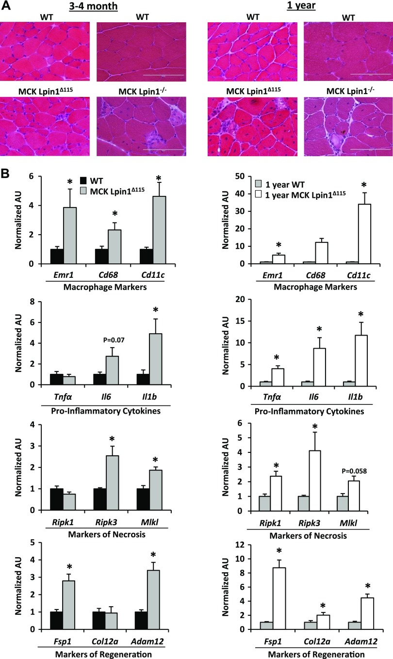
Figure 2.
Mice with muscle-specific Lpin1 deficiency exhibit active and progressive myopathy. A) H&E stains of skeletal muscle cross sections from MCK-Lpin1Δ115 and MCK-Lpin1−/− mice with respective age-matched WT controls at 3–4 mo and 1 yr old. Field width is ∼200 µm. B) Quantitative RT-PCR performed to quantify expression of the indicated genes from MCK-Lpin1Δ115 mice 3–4 mo and 1 yr old. Various markers of macrophage, proinflammatory cytokines, necrosis, and muscle regeneration were measured. Values were normalized (1.0) to age-matched WT mice, and data are shown as means ± sem. *P < 0.05 (Student’s t test) for differences in MCK-Lpin1Δ115 compared with age-matched WT mice; n = 7–9.

