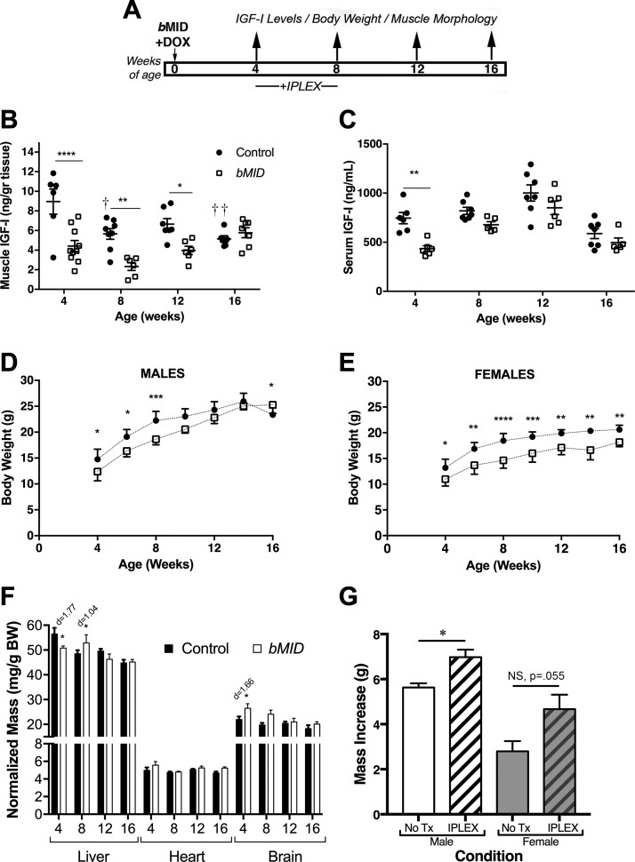
Figure 1.
Muscle Igf1 deletion leads to impaired body growth. A) Experimental design involving deletion of Igf1 at birth (bMID). B) IGF-I content of quadriceps was measured by ELISA in male and female bMID and control mice (4 wk, n = 6–10; 8 wk, n = 6–8, 12 wk, n = 6–7; and 16 wk, n = 6–9). bMID muscles had significantly lower IGF-I content at 4, 8, and 12 wk than age-matched control muscles. Control muscles exhibited an age-dependent decrease in Igf-I content. C) IGF-I levels quantified by ELISA in serum from male and female bMID and control mice (4 wk, n = 5–6; 8 wk, n = 5–6; 12 wk, n = 5–7; and 16 wk, n = 7–8). Circulating IGF-I was 40% lower in 4-wk-old bMID mice. D, E) Growth curves of male (n = 7–9) (D) and female (n = 5–10) (E) bMID and control mice from 4 to 16 wk of age show impaired growth of bMID mice until 8 wk in males and throughout the window of assessment for females. Male bMID mice became larger than control mice at 16 wk of age. F) Representative liver, heart, and brain dissections from 4-, 8-, 12-, and 16-wk-old bMID and control mice (n = 4–15), showed that loss of muscle IGF-I has only minimal effect on normalized mass of other tissues. G) Restoration of body growth after exogenous administration of mecasermin rinfabate. Histogram depicts the body mass increase of bMID males and females when injected with mecasermin rinfabate or vehicle between 4 and 8 wk of age. Data are means ± sem. Statistical analysis was performed with 2-way ANOVA, followed by Sidak’s multiple-comparisons test, or 2-tailed Student’s t text with effect size presented as Cohen’s d. *P < 0.05 (for bMID vs. control under same condition/treatment), **P < 0.01, ***P < 0.001, ****P < 0.0001, ††P < 0.01 (within-strain comparison).

