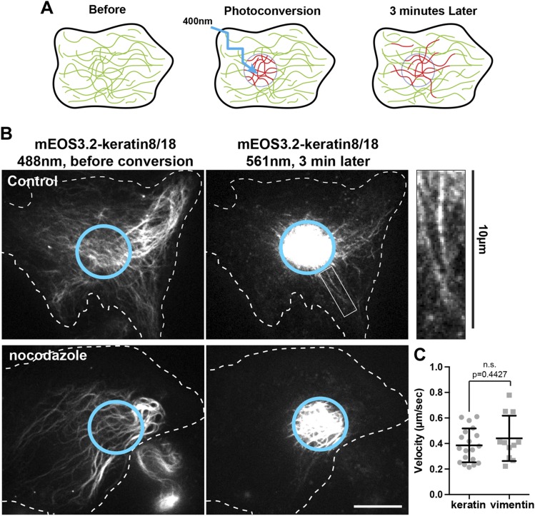
Figure 2.
Fully polymerized keratin filaments are transported along microtubules. A) Photoconversion strategy to study keratin filament transport. A subset of mEOS3.2-keratin8/18 filaments is photoconverted from green to red using 400 nm light. Transport of the photoconverted filament outside from the photoconverted zone (cyan circle) is monitored in the red channel during 3 min. B) Photoconversion of mEos3.2-keratin 8/18 in RPE cells using spinning disk confocal microscopy. The left panels show the keratin network in the green channel (488 nm) before conversion, and the right panels show the red channel (561 nm) 3 min after photoconversion. Several photoconverted filaments were present outside of the original photoconverted zone (cyan circle) in the control cells, whereas photoconverted filaments remained inside the initial zone in nocodazole-treated cells (10 µM for 3 h). Inset enlargement shows the presence of long filaments outside from the photoconversion area 3 min after photoconversion. Scale bar, 10 µm. C) The tip of at least 10 photoconverted filaments coming form 5 different cells has been tracked for a period of 15 to 30 s. The graph compares the velocity of vimentin and keratin filament in the control condition (mean ± sd). Significance was determined using the Mann-Whitney U test.

