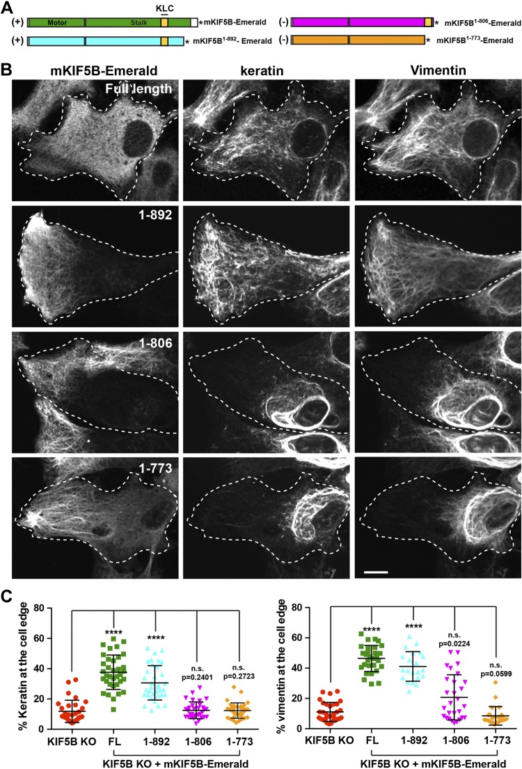
Figure 5.
Mouse KIF5B1–892 rescues IF distribution in KIF5B KO cells. A) Schematic representation of different truncations of the KIF5B tail. The mKIF5B constructs capable of rescuing keratin and vimentin distribution are marked by a (+); the one that does not is marked by a (−). The asterisk represents Emerald. B) Confocal microscopy imaging of keratin and vimentin immunostaining in RPE KIF5B KO cells (clone #4) after the expression of mKIF5B-Emerald (full length), mKIF5B1–892-Emerald (1–892), mKIF5B1–806-Emerald (1–806), or mKIF5B1–773-Emerald (1–773). C) Graphs show the percentage of vimentin (left) or keratin (right) at the cell edge (mean with sd, n > 30 cells). Data are representative of at least 2 independent experiments. Statistical significance was determined using the Mann-Whitney U test. ****P < 0.0001.

