Figure 2.
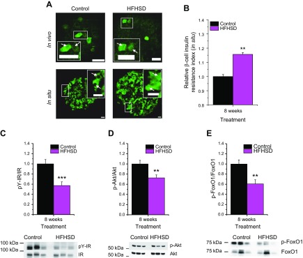
Verification of β-cell insulin resistance in HFHSD-fed mice. A) In vivo representative image of single-focal planes of engrafted islets transduced with βIRB in mice fed a control diet or HFHSD at 8 wk of diet treatment. In situ representative image of immunostaining of endogenous FoxO1 in situ in islets from pancreas sections from mice fed a control diet or HFHSD for 8 wk. Scale bars, 30 µm. B) Relative β-cell insulin resistance indicated by immunostaining of endogenous FoxO1 in situ in islets from pancreas sections from mice fed a control diet or HFHSD for 8 wk. C–E) Western blot quantification of pY-IR/IR, p-Akt/Akt, and p-FoxO1/FoxO1 in isolated islets from mice fed a control diet or HFHSD for 8 wk (n = 11–12). Representative Western blots for 3 control diet and HFHSD mice are shown. **P < 0.01, ***P < 0.001. Data are expressed as means ± sem. See also Supplemental Fig. S2.

