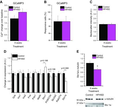
Figure 7.
Impaired insulin secretion of HFHSD mice is due to a defect downstream of glucose-stimulated Ca2+ influx. A) Ca2+ excursions upon glucose stimulus, as indicated by the GCaMP3 Ca2+ biosensor, obtained in vivo in islets from Ins-Cre:GCaMP3-mice transplanted into the ACE of B6 mice at 8 wk of an HFHSD (n = 6–9). B) Amount of glucose-responsive cells indicated by the GCaMP3 Ca2+ biosensor obtained in vivo in islets from Ins-Cre:GCaMP3-mice transplanted to the ACE of B6 mice at 8 wk of an HFHSD (n = 6–9). C) Backscatter intensity of islets transplanted to the ACE of B6 mice at 8 wk of an HFHSD shown in arbitrary units (A.U.) (n = 10). D) Gene expression analysis of isolated islets from mice fed a control diet or HFHSD for 8 wk shown in A.U. (n = 6 mice). E) Western blot quantification of syntaxin-1A in isolated islets from mice fed a control diet or HFHSD for 8 wk (n = 11–12). Representative Western blot of 3 control diet and HFHSD mice is shown. *P < 0.05, ***P < 0.001. Data are expressed as means ± sem. See also Supplemental Figs. S5 and S6.

