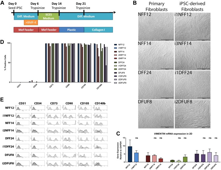
Figure 1.
Nondiabetic and diabetic iPSCs were differentiated into iPSC-derived fibroblasts. A) Schematic of the differentiation (Diff.) protocol. MEF, mouse embryonic fibroblast. B) iPSC-derived fibroblasts show typical fibroblast morphology by microscopy. C) iPSC-derived fibroblasts express mesenchymal marker, vimentin. D) iPSC-derived fibroblasts are positive for mesenchymal markers CD73, CD90, CD105, and CD140b and are negative for endothelial markers CD31 and CD34. E) Flow cytometry profiles of the mesenchymal and endothelial markers.

