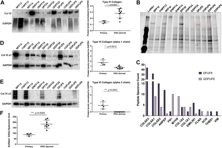
Figure 7.
In a 3D in vitro SA tissue model, iPSC-derived fibroblasts deposit ECMs that are distinct from primary fibroblasts. A) iPSC-derived fibroblasts are enriched in type IV collagen. B) Protein gel showing distinct band (red) of around 120 kDa present in primary and absent in iPSC-derived fibroblasts that was excised and analyzed using mass spectrometry analysis. C) Peptide spectrum counts of the identified proteins from the bands shown in B. D) Type VI collagen α-1 chain is down-regulated in iPSC-derived fibroblasts. E) Type VI collagen α-3 chain was not detected in iPSC-derived fibroblasts. F) Sulfated GAGs to HA ratio is higher in iPSC-derived fibroblasts compared with primary fibroblasts. Primary and iPSC-derived fibroblasts were compared using Student’s t tests; n = 2–4 of biologic replicates per each cell line. P > 0.05 (not significant), *P ≤ 0.05, **P ≤ 0.01, ***P ≤ 0.001, ****P ≤ 0.0001.

