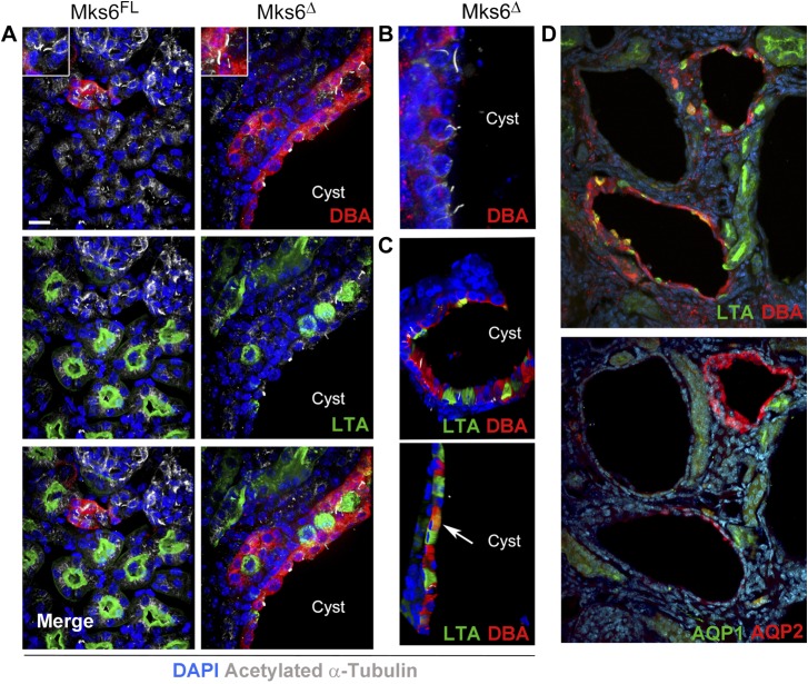
Figure 4.
Analysis of cystic tubules in Mks6Δ mutants. A, B) Immunofluorescence microscopy analysis for the presence of cilia, collecting and proximal tubules markers (acetylated α-tubulin in white, DBA in red, LTA in green). Original scale bar, 15 µm. A insets, B) Note the presence of cilia within a cyst in the Mks6Δ sample. C) Confocal microscopy-extended projections of mutant Mks6Δ tubules showing individual cysts lined by cells that express LTA, DBA, or both (arrow) within the same tubule. D) Immunofluorescence microscopy analysis for tubule markers (DBA and LTA) shows mixed cells in a Mks6Δ cyst, but Aquaporin 1 and 2 immunofluorescence (AQP1 and AQP2) shows cysts with uniform staining. Hoechst-stained nuclei are blue in all panels.

