Figure 5.
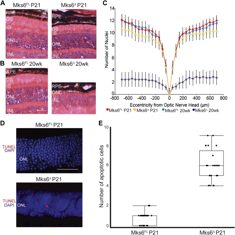
Retinal degeneration phenotype in Mks6Δ mutants. A, B) Hematoxylin and eosin staining of retina sections from Mks6FL and Mks6Δ mutants from juveniles (P21) and adults (20 wk). INL, inner nuclear retinal layer; ONL, outer nuclear retinal layer; RPE, retinal pigmented epithelia retina layer. Original scale bars, 50 µm. C) Morphometric analysis of nuclei counts at different distances from the optic nerve head using Student’s t test reveals only slight retinal degeneration in juveniles (P21) compared with statistically significant retinal degeneration in adults (20 wk). D) TUNEL staining images for apoptosis (red) in P21 retinas. DAPI-stained nuclei are blue. Original scale bar, 50 µm. E) Graph of TUNEL quantification in P21 retinas. Kruskal-Wallis nonparametric ANOVA with post hoc Mann-Whitney comparisons yielded P < 0.05; means ± sem, n = 6 per group.

