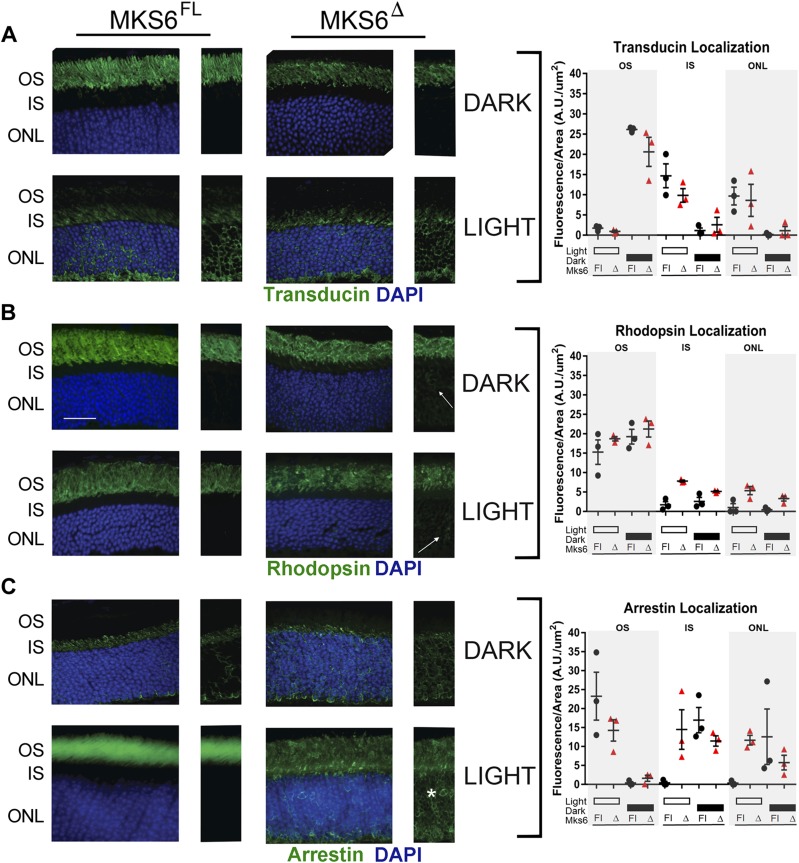
Figure 6.
Analysis of phototransduction protein localization in Mks6Δ retinas. A) Transducin staining (green) is similar between Mks6Δ mutants and Mks6FL controls in both the light and dark conditions. B) Rhodopsin staining (green) in both light- and dark-adapted conditions reveals slight mislocalization in the IS (arrows) in juvenile Mks6Δ mutants compared with Mks6FL controls. C) Arrestin staining (green) is present in the OS and is mislocalized to the IS and ONL in the light-adapted Mks6Δ mutant retinas (asterisk). One-way ANOVA was used for quantification of the average fluorescence intensity distribution indicated for each protein in the graphs (right). DAPI-stained nuclei are blue. Original scale bar, 25 µm.

