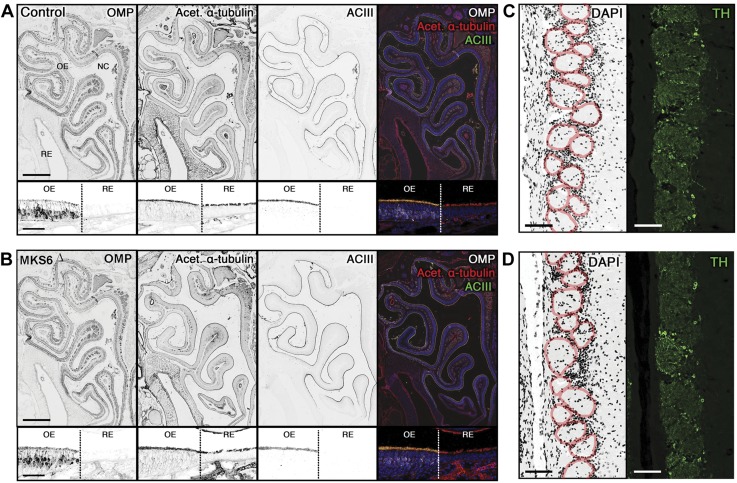
Figure 8.
Analysis of the olfactory system in Mks6Δ mutants. A, B) Global coronal section of the nasal cavity with the OE and RE from Mks6FL (Control) and mutant (Mks6Δ). Immunofluorescence for a marker for mature OMPs, and cilia markers (acetylated α-tubulin and ACIII). Closer examination of the OE and RE border (demarcated with dashed line) from Mks6FL and Mks6Δ mutant mice demonstrates the presence of olfactory sensory cilia and motile respiratory cilia in both mice. C, D) Coronal section of the OB medial surface from Mks6FL (Control) and mutant (Mks6Δ). DAPI stained with individual glomeruli (red outlines) and TH immunolabeling demonstrated no difference in glomerular size and trans-synaptic activation, respectively. Original scale bars, 500 µm (A, B, upper), 50 µm (A, B, lower); 100 µm (C, D), n = 3 per group.

