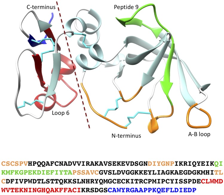
Figure 1.
The 3-dimensional structure of human TIMP2 and amino acid sequence showing the locations of the 2 domains and other regions discussed in the text. The representation was generated from pdb 1BR9 (105) using the University of California, San Francisco (UCSF) chimera package (106). Specific sections of the structure are colored as follows: reactive site, orange; peptide 9 (interaction with α3β1 integrin), green; loop 6 (interaction with IGF-1R), red; and C-terminal tail (MMP2–Hpx domain interaction), blue. Cystines and disulfide bonds are cyan, and the broken line marks the division between the N- and C-domains.

