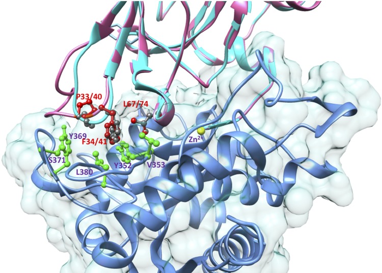
Figure 6.
A homology-based model of lamprey TIMPa superimposed on the crystallographic structure of the N-TIMP3/TACE complex, pdb 3CKI (58). The TIMPa model was generated by using the Swiss-Model Homology Modeling Server (108) based on structures of TIMP2 (pdb 1GXD and 2E2D). The structure was superimposed on the N-TIMP3 chain of 3CKI, and the image was generated by using the University of California, San Francisco (UCSF) chimera package (106). The backbones of TIMP3 and lamprey TIMPa are shown as cyan and pink ribbons, and the backbone of TACE is blue. The side chains of residues from TIMP3 are colored dark gray, and those from lamprey TIMPa are colored red. The labels for both are red. Side chains from TACE are colored green, and their labels are purple. The catalytic Zn2+ ion is shown as a yellow sphere.

