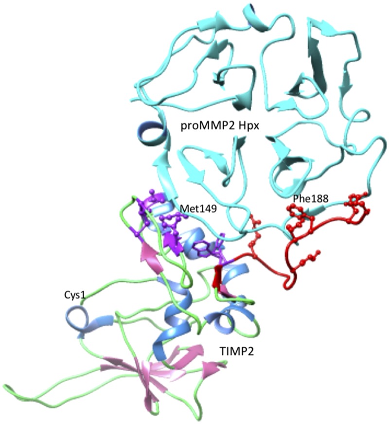
Figure 7.
Interactions between the TIMP2 and the Hpx domain of proMMP-2. The representation was generated from pdb 1GXD (105) by using the University of California, San Francisco (UCSF) chimera package (106). Specific parts of the complex structure are colored as follows: the chains of both proteins are displayed as ribbons, TIMP2 β-strands are pink, and α-helices are cornflower blue; the proMMP-2 Hpx domain is colored cyan. The side chains of TIMP2 residues that interact with the Hpx domain are displayed in ball and stick rendering. The group surrounding Met149 are colored purple, and the C-terminal tail and residues around Phe188 are colored red. The location of the N-terminal Cys of TIMP2 is labeled to show the location of the MMP-inhibitory region.

