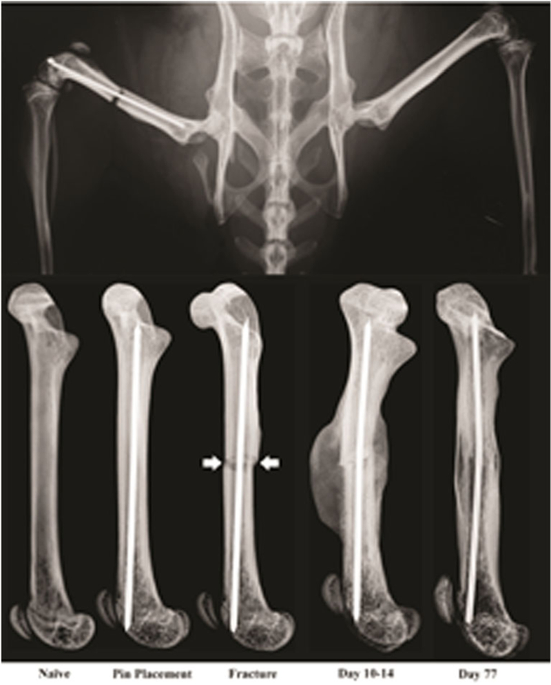
Fig. 1.

Representative radiographs showing the rodent model of bone fracture pain. Note that in the top image, a titanium pin has been placed in the intramedullary space of the right femur before the fracture (to stabilize the fractured bone) and a fracture has been made in the middle of the femur. Pain is immediately evident following bone fracture and with normal bone healing (callous formation, mineralization, resorption, and cortical union), the fracture pain subsides. These images are from a mouse but a nearly identical model has also been developed in rats
