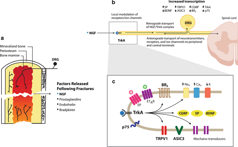
Fig. 3.

Schematic illustrating the sensory nerve fibers that innervate the femur and some of the factors that may contribute to fracture-induced skeletal pain. Sensory nerve fibers that innervate the bone (a) are generally mechanosensitive and are present in the periosteum (the thin cellular and fibrous sheath that surrounds the outer surface of the mineralized bone), cortical bone, and bone marrow. Following bone fracture, a variety of factors are released at the fracture site including nerve growth factor (NGF), prostaglandins, endothelins, and bradykinin which activate and/or sensitize neurons that convey information from the bone to the spinal cord (b) in that NGF binds to its cognate receptor tropomyosin receptor kinase A (TrkA) and the NGF/TrkA complex is then retrogradely transported to the cell body of the sensory neuron where it induces upregulation of a variety of neurotransmitters, receptors, and ion channels involved in detecting and transmitting noxious stimuli from the bone to the spinal cord and brain. In sensory nerve fibers at the fracture site (c), NGF also directly sensitizes a variety of receptors, ion channels, and mechanotransducers expressed by sensory nerve fibers that innervate the bone so that normally innocuous stimulation of the bone is now perceived as noxious stimuli
