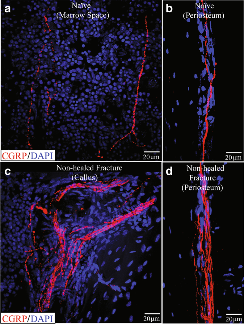
Fig. 4.

Sprouting of sensory nerve fibers at an unhealed fracture site 6 months following bone fracture where desired healing of the fracture has not occurred. In these confocal images, sensory nerve fibers are labeled with an antibody raised against calcitonin gene-related peptide (CGRP, red). Note that in the normal uninjured femur (a, b), there are a few new fibers in the bone marrow (a) and in the periosteum (b). However, in the non-healed fracture, there is a marked increase in the density of sensory nerve fibers in both the callous (c) and in the periosteum (D). This “hyper-innervation” of the bone at the unhealed fracture site is never observed in the normal femur and may contribute to the chronic limping and pain-related behaviors observed in animals with non-healed fractures
