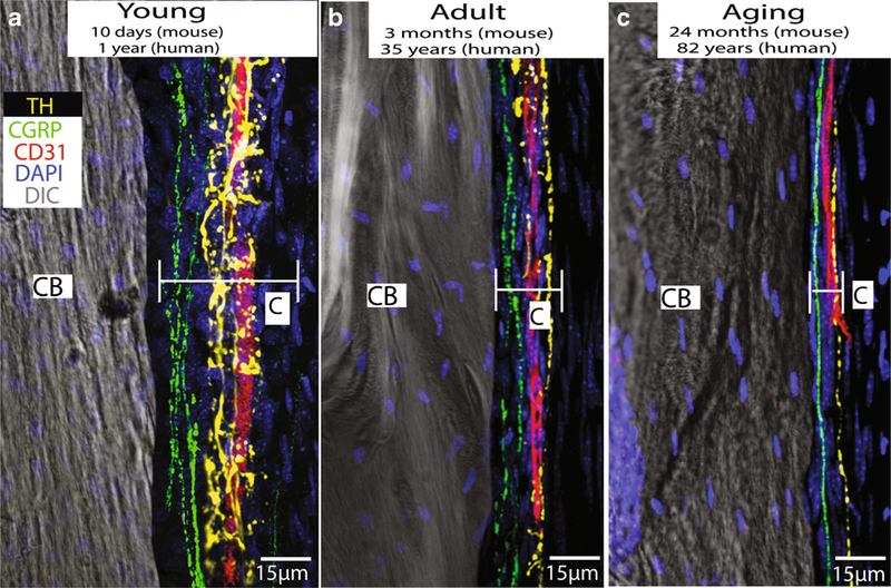
Fig. 5.

Images showing that sympathetic nerve fibers (yellow), sensory nerve fibers (green), and blood vessels (red) are present and have a similar organization in the periosteum of the young, adult, and aging mouse femur. The periosteum is a thin cellular and fibrous sheath that is tightly opposed to the outer surface of all bones of the body and probably plays a major role in detecting bone fracture and the generation of acute and chronic fracture pain. These data would suggest that even in the very young and very old, the periosteum is richly innervated by sensory and sympathetic nerve fibers and these nerve fibers probably play a significant role in the detection and signaling of fracture pain throughout the lifespan. In these images, blood vessels are labeled with an antibody raised against CD-31, primary afferent sensory nerve fibers are labeled with an antibody raised against calcitonin gene-related peptide (CGRP), and sympathetic nerve fibers are labeled with an antibody raised against tyrosine hydroxylase (TH)
