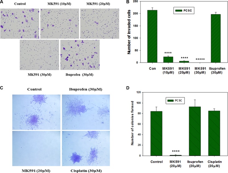
Figure 7. Effects of MK591 on in vitro invasion and soft-agar colony formation by PCSC.
In (A), invasive capabilities of PCS cells were assayed using matrigel-coated transwell chambers as described in the “Methods” section. After incubation, cells were fixed and stained with crystal violet. Pictures were taken with a Leica microscope at ×200. (B) Shows quantitative measurements of the number of invaded cells with or without drug treatment. Results represent mean values of individual data point ± standard deviation (n = 3). ****p = < 0.00005; *****p = < 0.000005. In (C), effects of MK591 on soft-agar colony formation by PCSC are shown. Cells were plated on soft-agar in complete medium and treated with drugs as indicated. After incubation for three weeks, cells were stained with crystal-violet and growing colonies were counted under microscope at ×150. Note: Dramatic inhibition was observed with MK591 treatment whereas the effects of ibuprofen and cisplatin were not distinguishable. In (D), results are shown quantitatively as mean values of each data point ± standard deviation (n = 3). ****p = < 0.00005.

