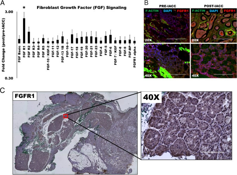
Figure 3. Fibroblast growth factor (FGF) signaling is upregulated following IACC in LGACC tumors.
(A) Bar graph of normalized quantitative fold expression of fibroblast growth factor (FGF) signaling family members in aggregate post/pre-IACC LGACC tumors identified in proteomic screening (statistically significant, p<0.05). (B) Immunofluorescence images of paired pre- and post-IACC LGACC samples probed for FGFR1 (red), and counterstained for filamentous actin (green), and nuclei (blue). (C) Representative immunohistochemical staining image for FGFR1 in a post-IACC LGACC tumor specimen. (micron bars: 20X=200 μm; 40X=100 μm).

