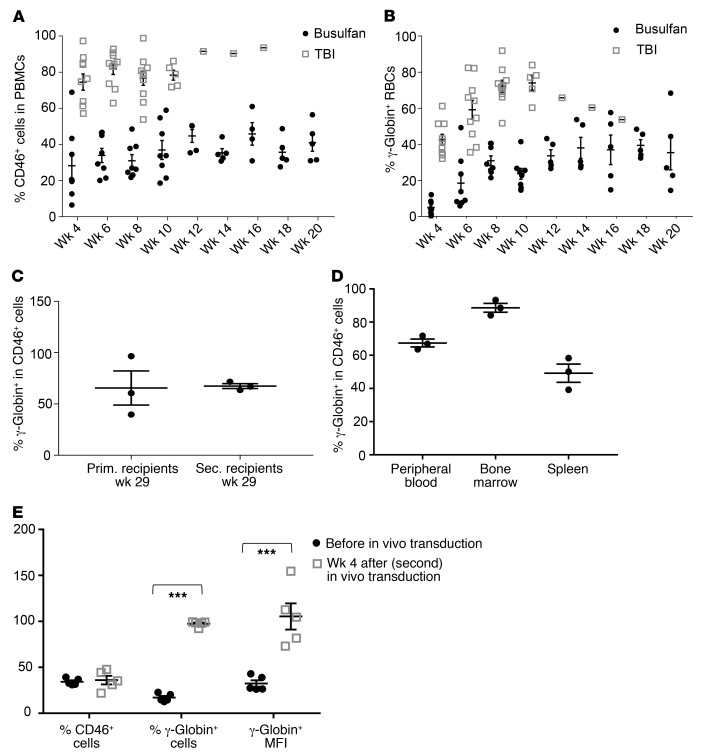
Figure 8. Analysis of secondary C57BL/6 recipients with transplanted bone marrow cells from treated CD46+/+/Hbbth-3 mice.
(A) Engraftment rates measured in the periphery based on the percentage of human CD46+ (hCD46+) cells in PBMCs after busulfan conditioning or total-body irradiation (TBI). (C57BL/6 recipients do not express hCD46.) (B) Percentage of human γ-globin–expressing peripheral blood RBCs. All mice received immunosuppression starting from week 4 after transplantation. (C) Percentage of γ-globin+ cells in hCD46+ (donor-derived) cells. (C and D) γ-Globin/CD46 expression in secondary C57BL/6 recipients at week 20 after transplant (busulfan preconditioning). CD46+ cells were immunomagnetically separated from the chimeric bone marrow of 3 representative secondary mice and analyzed for γ-globin expression by flow cytometry. Notably, unlike humans, huCD46tg mice express CD46 on RBCs. (C) γ-Globin/CD46 marking rates of primary and secondary recipients at sacrifice. (D) γ-Globin expression in CD46+-selected cells from the hematopoietic tissues of secondary recipients (week 20). Each symbol represents an individual animal. (E) γ-Globin expression in secondary recipients that received a new (second) round of HSPC mobilization/in vivo transduction (n = 5). Secondary recipients (busulfan-preconditioned) were analyzed for γ-globin and CD46 expression at week 20 after transplantation (“Before in vivo transduction”). These mice were then mobilized and transduced in vivo with the HDAd-γ-globin plus HDAd-SB vectors. Four weeks after in vivo transduction, mice were sacrificed and analyzed (“Week 4 after in vivo transduction”). ***P ≤ 0.00003. Statistical analyses were performed using 1-way ANOVA.

