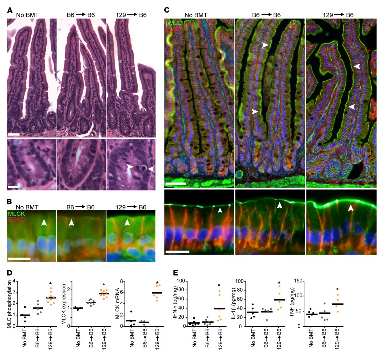
Figure 2. MLCK210 expression and activity as well as cytokines associated with MLCK210 upregulation are elevated in minor mismatch experimental GVHD.
B6 WT recipients were lethally irradiated followed by a syngeneic (B6) or allogeneic (129) BMT. Mice were sacrificed 14 days after BMT. (A) Representative histopathology of the small intestine. Arrowheads denote apoptotic epithelial cells. Scale bars: 50 μm (top), 10μm (bottom). (B) Jejunal segments were immunostained for MLCK210 (green) and E-cadherin (red). Arrowheads indicate the location of the perijunctional actomyosin ring. Images are representative of >3 independent experiments. Quantitative analysis is shown in D. Scale bar: 10 μm. (C) Jejunal segments were immunostained for phosphorylated myosin light chain (pMLC, green) and E-cadherin (red). Arrowheads indicate the location of the perijunctional actomyosin ring. Images are representative of >3 independent experiments. Quantitative analysis is shown in D. Scale bars: 50 μm (top), 10 μm (bottom). (D) MLCK210 expression and MLC phosphorylation were determined morphometrically. Each point represents an average of 4 fields from one segment of tissue from a single mouse. Two segments were analyzed per mouse. Data are normalized to the mean of mice that did not receive BMT. MLCK210 mRNA was determined by quantitative PCR (qPCR) in purified epithelial cells. Each point represents an individual mouse. Data are normalized to the mean of mice that did not receive BMT. *P < 0.05, 2-tailed t test (B6→WT vs. 129→WT). (E) Jejunal cytokines were determined by ELISA. Each point represents an individual mouse. *P < 0.05, 2-tailed t test (B6→WT vs. 129→WT).

