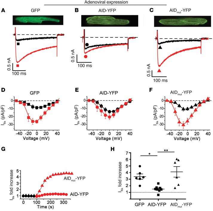
Figure 3. β-less WT endogenous CaV1.2 channels are not stimulated by PKA.
(A–C) Adenovirus-induced GFP, AID-YFP, and AID-mutant YFP expression in cultured guinea pig ventricular myocytes. Top: exemplar confocal images from guinea pig cardiomyocytes expressing GFP, AID-YFP peptide, or AID-mutant YFP peptide. Bottom: exemplar whole-cell Ba2+ currents from GFP and YFP-expressing guinea pig ventricular cardiomyocytes before (black trace) and after (red trace) application of 1 μM forskolin. (D–F) Current-voltage relationships from GFP, AID-YFP, and AID-mutant YFP–expressing cardiomyocytes before (black) and after (red) superfusion of 1 μM forskolin. (G) Representative diary plot showing time course of forskolin-induced increase in CaV1.2 current. (H) Forskolin-induced increase in CaV1.2 current. *P < 0.05, **P < 0.01 by 1-way ANOVA and Tukey’s multiple comparison test.

