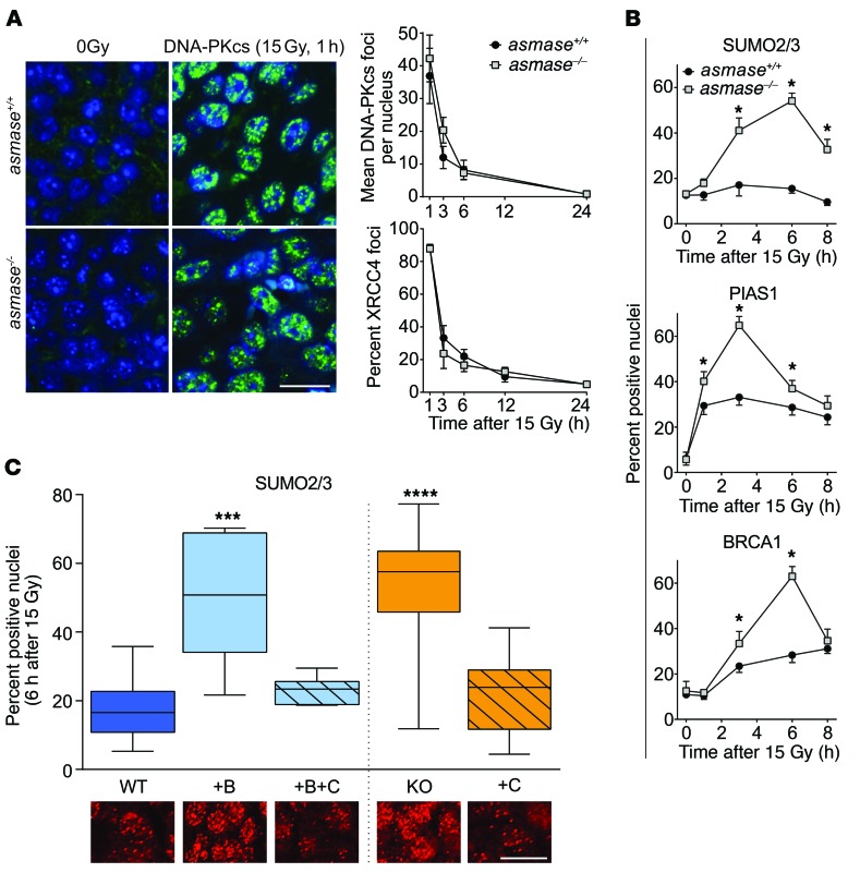
Figure 3. I/R confers epigenetic HDR loss of function.
Foci were scored as in Figure 2 in asmase+/+ (WT) or asmase–/– (KO) hosts. (A) Canonical NHEJ is insensitive to SDRT-I/R. Left: Representative DNA-PKcs foci in MCA/129 fibrosarcomas 1 hour after 15 Gy SDRT. Right: Time-dependent change in DNA-PKcs and XRCC4 foci in SDRT-treated B16F1 melanomas. Scale bar: 20 μm. Data represent mean ± SEM collated from 2–4 independent experiments per panel of 3 mice per group. P > 0.05, WT vs. KO. (B) Time course of SUMO2/3, PIAS1, and BRCA1 focus accrual/resolution after 15 Gy SDRT in HCT116 xenografts. *P < 0.05, WT vs. KO unpaired t test. Data represent mean ± SEM collated from 2–4 independent experiments per panel of 3 mice per group. *P < 0.05, WT vs. KO. (C) Effects of SDRT-I/R injury (WT, WT+B+C, KO+C) versus SDRT-inert (WT+B, KO) settings on SUMO2/3 foci formation in HCT116 tumor xenografts at 6 hours after 15 Gy SDRT. BQ-123 (designated B), when used, was injected i.p. 30 minutes before SDRT, while mechanical percutaneous clamp (designated C) of large tumor-feeding vessels was used immediately after SDRT. Data represent median ± IQR percent foci-positive nuclei in tumor-derived histological specimens from 2–4 mice each, scoring a total of 2 × 10³ to 7 × 10³ HCT116 cells. ***P < 0.001, ****P < 0.0001 vs. WT, with Bonferroni correction (threshold: α = 0.05/4 = 0.0125). Inset shows representative SUMO2/3 focus images in respective SDRT-I/R–conditioned and I/R-inert. Scale bar: 20 μm.

