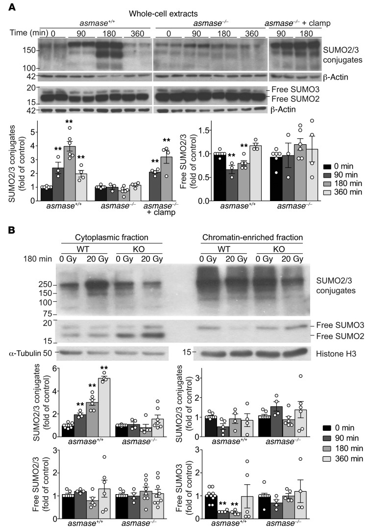
Figure 4. I/R disrupts SUMO2/3 function in tumors exposed to SDRT.
Western blot (WB) analysis of tumor extracts using rabbit polyclonal anti-SUMO2/3 antibody, quantified by densitometry relative to loading controls. (A) Whole-cell extracts from MCA/129 fibrosarcoma in asmase+/+ and asmase–/– hosts after 20 Gy SDRT. Top panels show representative WBs, and bottom panels quantify high-MW SUMO2/3 conjugates (>75 kDa) and free SUMO2/3. (B) Representative WBs of high-MW SUMO2/3 conjugates (>75 kDa) and free SUMO2/3 in cytoplasmic (left) and chromatin-bound (right) fractions isolated from MCA/129 fibrosarcomas in asmase+/+ and asmase–/– hosts at 3 hours after 20 Gy SDRT. Bottom panels show quantitative analysis of specimens at different times after 20 Gy. (A and B) Data represent mean ± SEM of at least 3 independent experiments of 2 mice per group. **P < 0.01 vs. 0min, Bonferroni correction (threshold: α = 0.05/3 = 0.017) .

