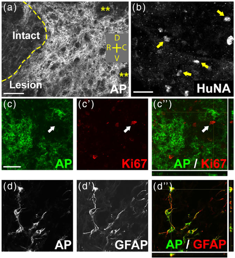
FIGURE 2.
GRP transplants survived long term and efficiently differentiated into astrocytes in the injured spinal cord. AP-labeled GRP-derived cells robustly survived within the lesion site for at least 5 weeks post-transplantation (a). Yellow asterisks in (a) denote areas within the lesion devoid of AP+ cells. Orientation: D = dorsal, V = ventral, R = rostral, C = caudal. HuNA+ fibroblast transplants survived in the lesion site until the 5-week point of sacrifice (b). arrows in (b) denote HuNA+ cells. At 5 weeks post-injection, AP+ cells did not continue to express the proliferation marker Ki67 (c–c”). Arrows in (c–c”) denote a Ki67+ cell that was not AP+. The vast majority of AP+ cells at 6 weeks post-transplantation differentiated into GFAP+ astrocytes (d–d”). Scale bar: 50 μm

