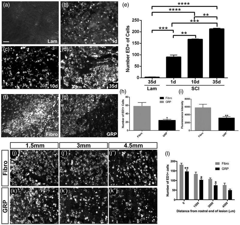
FIGURE 9.
GRPs significantly reduced the macrophage response both within the lesion site and in the intact caudal spinal cord surrounding PhMNs. There were almost no ED1+ cells in the intact C2 spinal cord in laminectomy-only control rats (a). There was a substantial increase in ED1+ cell counts by 1 day posthemisection (b), and these counts progressively increased out to 10 days (c) and 35 days (d) post-SCI (quantification in e). Compared with fibroblasts (f), GRPs (g) significantly reduced the number of ED1+ cells (h) and the intensity of ED1 immunostaining (i) in the lesion site at 5 weeks post-SCI. Compared with fibroblasts (j–j”), GRPs reduced numbers of ED1+ cells in the ipsilateral C3–C5 ventral horn (k–k”) at 5 weeks posthemisection (l). Scale bar: 50 μm

