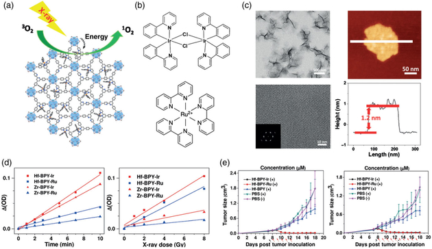
FIGURE 5.
Nanoscale metal organic layers for X-PDT. (a) Schematic for Hf-based nMOL X-PDT agent. (b) Photosensitizers. Top: Ir[2,2′-bipyridine (2-phenylpyridine)2]+. Bottom: [Ru(2,2′-bipyridine)3]2+ c) physical characterization. Top left: TEM nMOL bottom left: HRTEM image, inset: FFT pattern. Top right: Tapping-mode AFM topography. Bottom right: Height profile along the white line. d) SOSG assay results. Samples were treated by 225 kVp irradiation. (e) in vivo therapeutic effect of nMOL (left:CT26, right:MC38). (Reprinted with permission from Lan et al. (2017). Copyright 2017 John Wiley and Sons)

