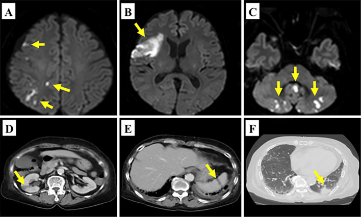
Figure 1.
Diffusion-weighted magnetic resonance imaging showed multiple high-intensity signal lesions in the right frontal lobe, temporal lobe and cerebellum (A-C: yellow arrows). Contrast-enhanced computed tomography showed low-density areas in the right kidney (D: yellow arrow) and spleen (E: yellow arrow). Lung tumor in the left inferior lobe was incidentally detected (F: yellow arrow).

